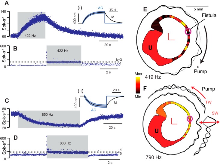
Fig. 9.
Auditory frequency responses with dehiscence or fistula. A–D: single-unit afferent neuron responses to auditory frequency stimuli in an animal model of dehiscence. A and B: example afferent neuron excited by 422-Hz auditory frequency stimulation through nonlinear endolymph pumping causing slow buildup of discharge rate (A) and another afferent in the same animal phase locking action potential firing to the auditory frequency stimulus at winding ratios (B), k = 3,4,5. Insets i and ii: illustration of the slow component of cupula displacement (M, black) evoked by the auditory frequency driven endolymph pumping and cycle by cycle vibration around the deflected position (AC, blue). C and D: example afferent neuron inhibited by endolymph pumping for a 850-Hz stimulus (C) and another afferent in the same animal phase locking action potential at winding ratios (D), k = 4.5. E and F: computational model of a human semicircular canal showing slowly developing pressure distribution (yellow: high; red: zero; and black: low) evoked by auditory frequency stimulation at 419 Hz (E) and 790 Hz (F). Waves travel along the membranous labyrinth away from the site of the fistula causing vibration of hair bundles at the stimulus frequency and pumping of endolymph in either direction, ampullofugal for 419 Hz (E) and ampullopetal for 790 Hz (F). SW, standing waves; TW, traveling waves. [Based on Iversen et al. (2018).]

