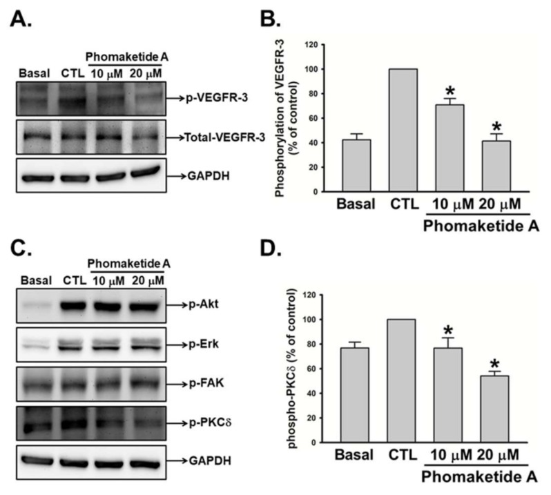
Figure 3.
Modulation of phomaketide A on VEGFR-3 and downstream signaling pathways in human LECs. (A and C) Quiescent LECs were treated with or without EGM-2MV medium in the absence (CTL) or presence of phomaketide A for 5–10 min. The phosphorylation of VEGFR-3, Akt, Erk, FAK, and PKCδ were determined by Western blot analysis (N = 5–7). The quantitative densitometry of the relative levels of phosphorylated VEGFR-3 and PKCδ were measured by Image-Pro Plus processing software (B and D). Data are expressed as the mean ± SEM. * p < 0.05 compared with the control (CTL) group.

