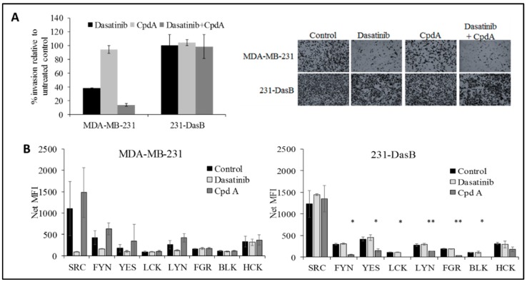
Figure 4.
(A) Invasion assays in MDA-MB-231 and 231-DasB cells with/without dasatinib (100 nM) and/or CpdA (5 μM); (B) phosphorylation of SFKs in MDA-MB-231 and 231-DasB in response to dasatinib 100 nM and CpdA (5 µM) where NET MFI is net median fluorescence intensity. * p < 0.05, ** p < 0.01. Error bars represent the standard deviations of triplicate independent experiments. p values were calculated using the Student’s t-test.

