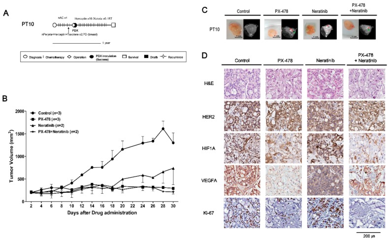
Figure 5.
In vivo efficacy of PX-478 and neratinib against PDX models from PT10 (HR−/HER2+ subtype). (A) PT10 treatment history. See text for details. Scale bar, one year. (B) Tumor volumes (F3) were determined in female mice (n = 2–3) given PX-478 (10 mg/kg), neratinib (20 mg/kg), or their combination three times a week for 30 days by peroral administration; mice administered saline served as controls. Tumor volumes at 30 days are presented as means ± SD (p ≤ 0.05 for control vs. PX-478 and control vs. PX-478 + neratinib; p = 0.32 for control vs. neratinib; unpaired t-test). (C) MRI images of tumors at 30 days after administration of drugs. Red arrows represent necrosis and green arrows are traces of blood flow. (D) H&E staining and immunohistochemical analyses of HER2, Ki-67, VEGFA and HIF1A in PDX tumors from PT10 after treatment with drugs. Scale bar, 200 μm.

