Figure 4.
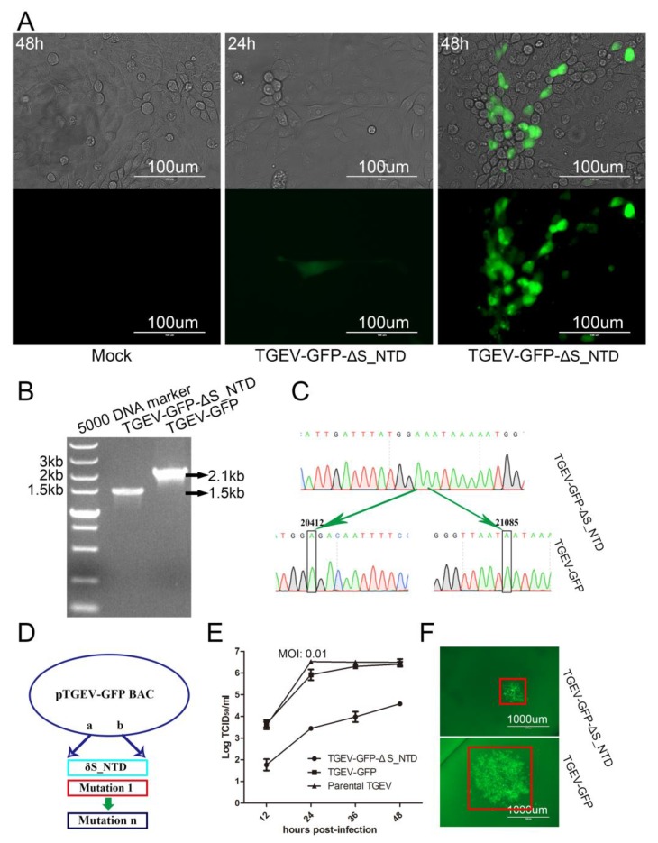
Rescue of TGEV-GFP-ΔS_NTD infectious clone in PK-15 cells. (A) CPE or fluorescence microscopy of PK-15 cells infected with the recombinant virus TGEV-GFP-ΔS_NTD or mock infected at 24 and 48 h post-transfection. (B) Electrophoresis detection of recombinant TGEV-GFP and TGEV-GFP-ΔS_NTD by RT-PCR using the primers rec-672SF and rec-672SR; (C) Sequence analysis of the targeted mutation area between recombinant TGEV-GFP and TGEV-GFP-ΔS_NTD by RT-PCR sequencing. (D) Model for the simultaneous construction of numerous infectious clones including S_NTD224. (E) Growth curves with the wild-type viruses TGEV-GFP and TGEV-GFP-ΔS_NTD with an original MOI of 0.01. (F) Viral fluorescent plaques between recombinant TGEV-GFP and TGEV-GFP-ΔS_NTD. The red box represents the size of the viral fluorescent plaques.

