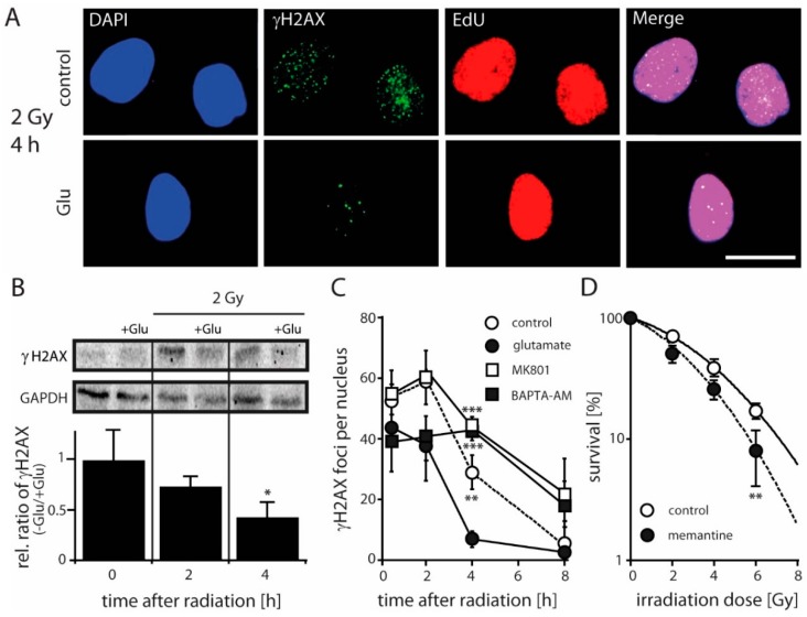
Figure 3.
Effect of glutamate on DSB repair and clonogenic survival of LN229 cells. (A) Immunofluorescence of γH2AX, EdU and DAPI from Glu-treated and untreated cells 4 h after irradiation (2 Gy). Note that addition of Glu (1 mM) resulted in a significant decrease in DSB levels in EdU-positive cells compared to mock-irradiated control cells from 29 ± 5 to 7 ± 2 foci (p < 0.01; n = 3; Student’s t-test). Scale bar 20 µM. (B) γH2AX protein levels and their relative ratio (normalized to GAPDH expression) in the absence and presence of Glu (1 mM) before and after IR with a dose of 2 Gy detected by western blotting at the indicated time points (* p < 0.05; n = 3; Student’s t-test). (C) Repair kinetics of γH2AX foci in S/G2 phase cells after IR (2 Gy) under control conditions, in the presence of Glu (1 mM), MK801 (10 µM) and BAPTA-AM (3 µM). The difference between Glu-treated/untreated or MK801 and BAPTA-AM treated cells at the time point of 4 h is determined by one-way ANOVA followed by Bonferroni’s post-hoc test, ** p < 0.05, *** p < 0.001. (D) Colony formation of cells treated with memantine (50 µM) upon IR. The calculated radiation-induced cytotoxicity enhancement factor at 2 Gy is 1.4. Data are given as means ± SD of three independent experiments. The significant difference between treated and untreated cells is determined by Student’s t-test (** p < 0.01).

