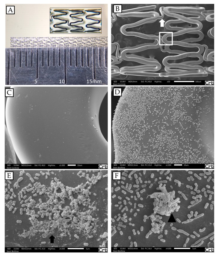
Figure 2.
A. baumannii biofilm formations on fragments of cobalt–chromium vascular stents analyzed by SEM. (A) Coronary stent photography; the fragments used in the experiments were 6 mm long. (B) Architecture of the cobalt–chromium vascular stent, presenting two cells, connected by a link (arrow). (C) Stent structure enlargement of box in B. (D and E) Stent after incubation with A. baumannii AB 72 for 24 h under conditions for biofilm formation (arrow). (F) Protuberant bacterial accumulation, suggestive of initial biofilm formation (arrow head). Magnifications: (B) 40×; (C) and (D) 1000×; (E) and (F) 4000×.

