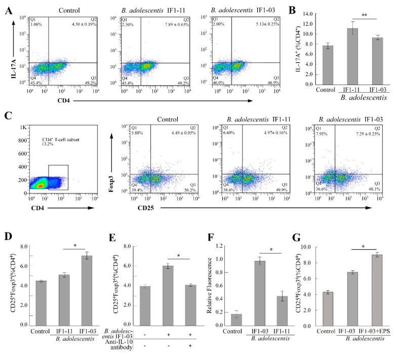
Figure 2.
Effect of two B. adolescentis strains on differentiation and proliferation of Th17/Treg cells in coculture with splenocytes. Splenocytes isolated from BALB/c mice were stimulated with B. adolescentis IF1-11, B. adolescentis IF1-03 or left untreated (control) for 72 h. (A) Representative flow cytometric dot plot and (B) proportion of Th17 cells (CD4+IL-17A+) in spleen lymphocytes. (C) Representative flow cytometric dot plot and proportion of Treg cells (CD4+CD25+Foxp3+) to CD4+ T cells. SSC-H (side scatter-height) the basic parameter indicating cell internal complexity, FL2-H (Foxp3, PE), FL3-H (CD4, PerCp—Cy5.5), and FL4-H (CD25, APC) emitted fluorescence signal height of different fluorescent dyes, by flow cytometry analysis. (D) Proportion of Treg cells (CD4+CD25+Foxp3+) in spleen lymphocytes stimulated with strain IF1-11 or IF1-03. (E) Effect of the addition of anti-IL-10 antibody on the induction of Treg cells induced by B. adolescentis IF1-03. (F) Amount of EPSs in B. adolescentis IF1-03 and B. adolescentis IF1-11. (G) Proportion of Treg cells (CD4+CD25+Foxp3+) in splenocytes stimulated with B. adolescentis IF1-03 cells alone or in the presence of IF1-03 EPS for 72 h. Data are shown as mean ± SD for five independent experiments and the significances between two B. adolescentis strains (A–F), with or without the addition of B. adolescentis IF1-03 EPS (G) were analyzed by one-way ANOVA, (* p < 0.05, ** p < 0.01).

