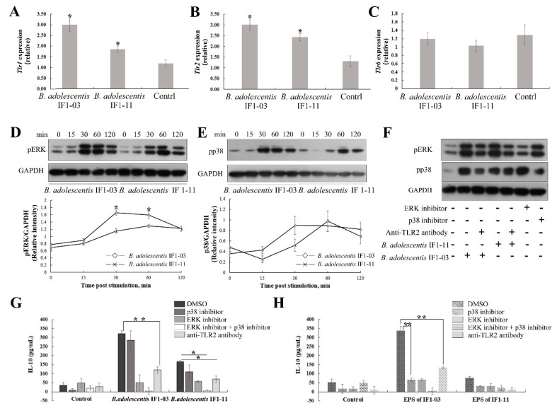
Figure 5.
Activation of TLR2 and subsequent ERK/p38- MAPK signaling in RAW264.7 macrophages after stimulation with two bifidobacterial strains. Total RNA of macrophages stimulated with B. adolescentis IF1-11 and B. adolescentis IF1-03, and control macrophages, was isolated at 4 h for cDNA synthesis and assay. Expression of Tlr1 (A), Tlr2 (B) and Tlr6 (C) was analyzed by qRT-PCR. Western blot analysis of the phosphorylated ERK (pERK) (D) and phosphorylated p38 (pp38) (E) in treated macrophages at the indicated times. Western blot analysis of pERK and pp38 of B. adolescentis IF1-03 and IF1-11-stimulated macrophages in the presence of anti-mouse TLR2 antibody, and ERK/p38-MAPK signal pathway inhibitors (F). IL-10 synthesis and secretion in macrophages stimulated with B. adolescentis IF1-11 and IF1-03 strains, in the presence of anti-mouse TLR2 antibody and specific chemical inhibitors of ERK (PD98059, 10 μM) and p38 (SB203580, 10 μM) phosphorylation (G). EPSs of different strains stimulate IL-10 production of macrophages in the presence of TLR2 antibody and inhibitors, respectively. IL-10 concentration in supernatants of cell cultures was determined by ELISA (H). Data are shown as mean ± SD for three independent experiments, statistically significant difference is indicated (* p < 0.05, ** p < 0.01).

