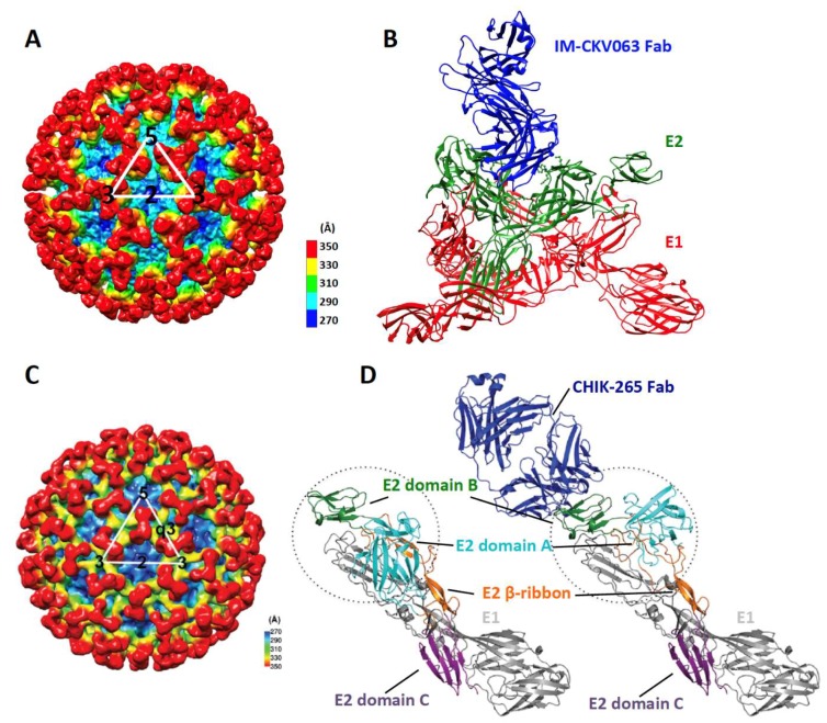
Figure 3.
Binding of IM-CKV063 and CHK-265 to CHIKV. (A) CryoEM reconstruction of CHIK VLP in complex with IM-CKV063 Fab fragment (EMD-6457); (B) Structure of the trimeric spike bound with IM-CKV063 Fab as observed in (A), showing IM-CKV063 crosslinks two neighboring E2 molecules within one trimeric spike. E1, E2 and Fab are shown in red, green and blue, respectively. (C) CryoEM reconstruction of CHIK vaccine strain 181/25 in complex with CHIK-265 Fab fragment; (D) (Left) Structure of the E1-E2 heterodimer (PDB:3N42); (Right) Structure of the E1-E2 heterodimer bound with CHIK-265 Fab as observed in (C), showing the repositioning of E2 domain A upon CHIK-265 binding to E2 domain B. The E2 domain A and domain B are circled, showing the difference in their conformations with and without Fab CHIK-265 binding (adapted from [6] with permission).

