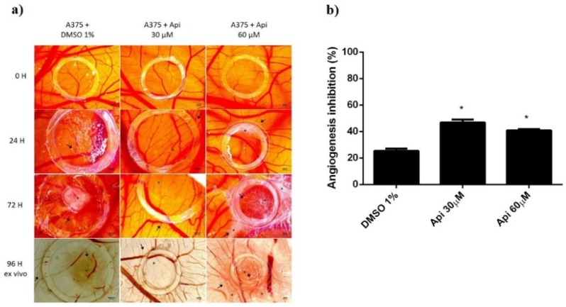
Figure 12.
A375 melanoma cells on CAMs treated with Api: (a) Stereomicroscopic in vivo images of the areas previously inoculated with melanoma cells and treated with Api 30 and 60 µM and with DMSO 1% as solvent control; initially—0 h, after 24 h, after 72 h, and after 96 h, ex vivo, after membranes biopsies were obtained; (b) the angiogenic inhibition % in A375 melanoma cells environment, at 48 h, for Api 30 µM and 60 µM compared to DMSO 1%. * p < 0.05.

