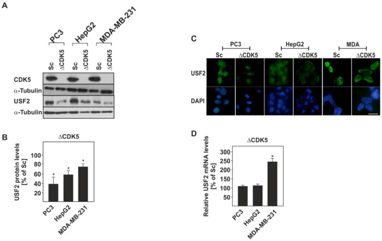
Figure 3.
Lack of CDK5 alters USF2 protein and mRNA levels in PC3, HepG2, and MDA-MB-231 cancer cell lines. (A) Representative western blot of USF2 protein levels. (B) Quantification of USF2 protein levels. USF2 levels in all Sc cells were set to 100%. Values represent means ± SD of at least three independent experiments. (C) Representative fluorescence images from the respective Sc and ΔCDK5 PC3, HepG2 and MDA-MB-231 cancer cell lines probed with an antibody against USF2 and stained with DAPI. Bar, 10 µm. (D) USF2 mRNA levels were measured by quantitative RT-PCR and mRNA levels in all Sc cells were set to 100%. Values represent means ± SD of at least three independent experiments. Statistics: Student’s t-test for paired values; *, significant difference Sc versus CDK5 knockout cells; p ≤ 0.05.

