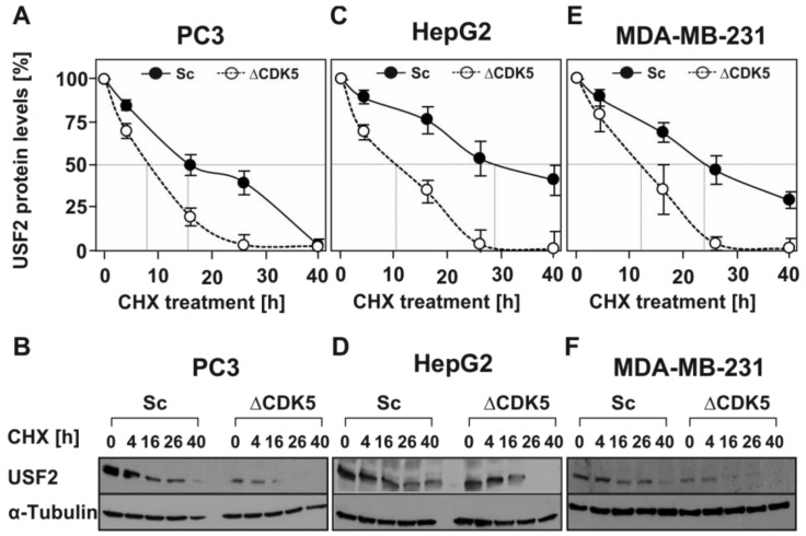
Figure 4.
Absence of CDK5 reduces the half-life of USF2. (A,C,E) Respective Sc and ΔCDK5 cells were treated with 10 µg/mL CHX for the indicated time periods. Proteins were isolated, separated by SDS-PAGE and detected by western blotting. Protein levels were quantified and the relative protein level of USF2 was blotted against the duration of CHX treatment for estimation of the half-life. The dashed line indicates the USF2 half-life where 50% of the USF2 protein level was reached. (B,D,F) Representative western blots. 50 µg of protein was probed with an antibody against USF2 and α-tubulin.

