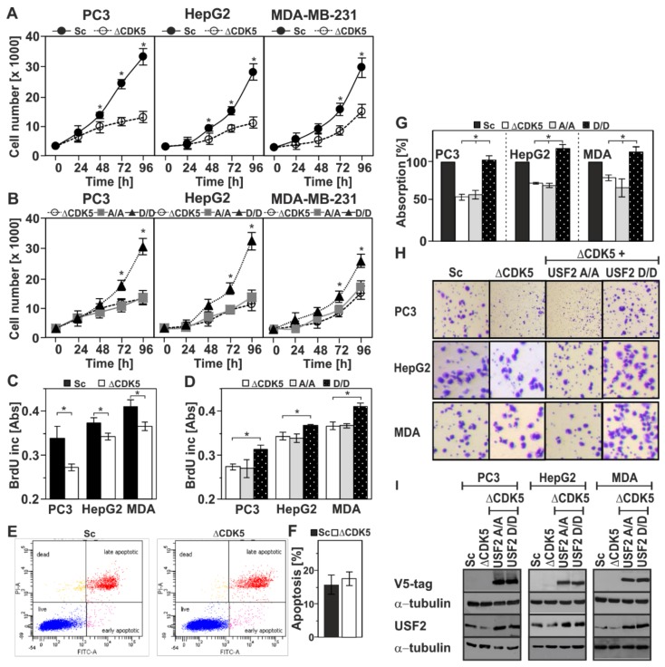
Figure 5.
Phosphorylation of USF2 by CDK5 affects cell proliferation and migration. Cellular proliferation was monitored by cell number (A,B) and BrdU incorporation (C,D) in Sc and ΔCDK5 cells (A,C) and in ΔCDK5 cells expressing either non-phosphorylatable USF2-S155A/S222A or phosphomimetic USF2-S155D/S222D double mutants (B,D). (E,F) Apoptosis in Sc and ΔCDK5 PC3 cells was assessed by Anexin-V/PI staining and analyzed by flow cytometry; (G) Migration was assessed by crystal violet staining. The values represent the absorbance of crystal violet at 595 nm. *, significant difference, p ≤ 0.05. (H) Representative images of a Transwell migration assay. (I) The expression of USF2 was controlled by western blot with an antibody against V5-tag and USF2; α-tubulin served as a loading control.

