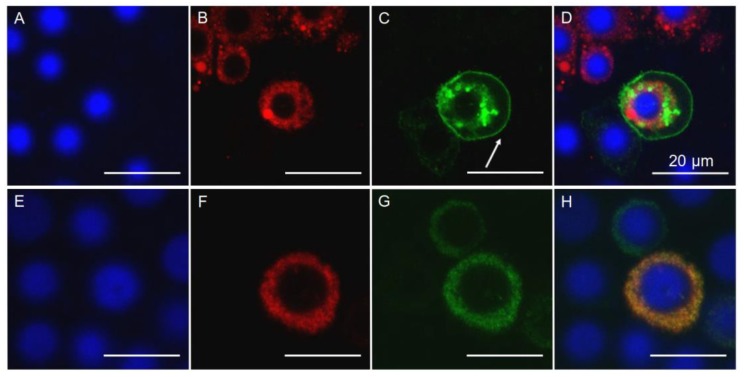
Figure 2.
Cellular localization of PgCad1 proteins within Hi5 cells. Hi5 cells transfected with pIE2-sPgCad1-GFP (A–D) and pIE2-r16PgCad1-GFP (E–H). Nuclei stained with Hoechst 3342 are shown in blue, dsRED-labeled endoplasmic reticulum is shown in red, and GFP-labeled PgCad1 fusion proteins are shown in green. Superimposed images from (A–C) are shown in (D) and from (E–G) in (H). The arrow in (C) indicates the cell membrane. Bar = 20 μm.

