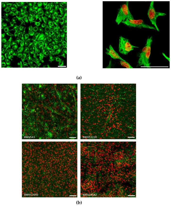
Figure 3.
Vimentin staining in human GBM: (a) U251 cells grown as monolayers (left panel, field of view 400 by 400 μm, scale 50 µm) and high magnification (right, scale bar = 50 µm) of single cells, vimentin staining is presented here as a high dynamic range (HDR) image showing the perinuclear vimentin-rich region and fine network of filaments. (b) Surgically obtained human tumor specimens were stained for vimentin, field of view 400 by 400 µm, scale 50 µm.

