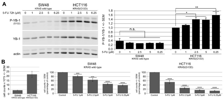
Figure 2.
5-FU induces Y-box binding protein 1 (YB-1) phosphorylation at S102 in KRAS(G13D)-mutated HCT116 cells but not in KRAS wild-type SW48 cells. (A) KRAS wild-type SW48 and KRAS(G13D)-mutated HCT116 cells were treated with increasing concentrations of 5-FU for 72 h. Thereafter, protein samples were isolated, and phosphorylation of YB-1 was analyzed by Western blotting using a phospho-specific antibody. Actin was detected as the loading control. The histogram represents the mean ratio of phosphorylated YB-1 (P-YB-1)/YB-1 from three independent experiments normalized to untreated HCT116 control cells. (B) A proliferation assay was performed following the same treatment conditions. Histograms indicate the mean number of cells after treatment with the indicated concentrations of 5-FU normalized to the control condition in each cell line (9 data points from three biologically independent experiments). Asterisks indicate a significant antiproliferative effect of 5-FU as analyzed by Student’s t-test (* p ≤ 0.05, ** p ≤ 0.01, *** p ≤ 0.001, and **** p ≤ 0.0001; n.s.: nonsignificant). (B, left part) Comparison of absolute cell counts of control conditions in SW48 and HCT116 cells.

