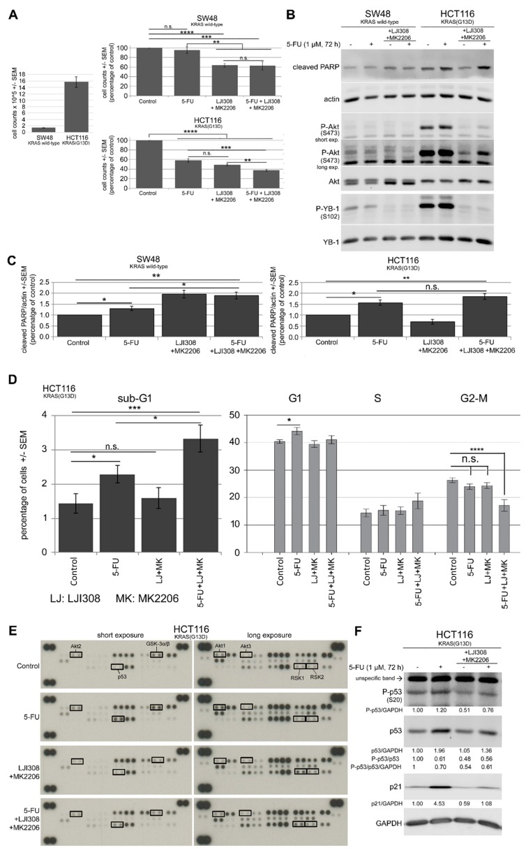
Figure 5.
Dual targeting of the RSK/YB-1 pathway and Akt enhances the antiproliferative effect of 5-FU in KRAS(G13D)-mutated HCT116 cells but not in KRAS wild-type SW48 cells by stimulating apoptosis. KRAS wild-type SW48 and KRAS(G13D)-mutated HCT116 cells were treated with 5-FU (1 µM), a combination of the RSK and Akt inhibitors (5 µM LJI308 for SW48 and 10 µM LJI308 for HCT116 cells; 5 µM MK2206) or a combination of the inhibitors with 5-FU for 72 h. (A) A proliferation assay was performed as described in the Materials and Methods section, and the mean value of 9 data points from three independent experiments is shown. (B) After the indicated treatments, protein samples were isolated, and the levels of P-YB-1, P-Akt, YB-1, Akt1 and cleaved PARP were determined by Western blotting. Actin was detected as a loading control. (C) The ratio of cleaved PARP to actin was evaluated by densitometry from three independent experiments and graphed. (D) The percentage of sub-G1, G1, S, and G2/M cells after the described treatment was investigated by fluorescence-activated cell sorting (FACS) analysis, as described in the Materials and Methods section. Data represent the mean value of 10 data points from four independent experiments for sub-G1 cells and 9 data points from three independent experiments for G1, S and G2-M phases. Asterisks indicate significant differences between the indicated groups (* p ≤ 0.05, ** p ≤ 0.01, *** p ≤ 0.001 and **** p ≤ 0.0001). (E) A phospho-mitogen-activated protein kinase (MAPK) array was performed in HCT116 cells according to the manufacturer’s protocol after the described treatments. (F) Western blot analysis was performed to analyze P-p53 (S20), p53, p21 and GAPDH in the protein samples, which were applied to the phospho-MAPK array. Densitometry values below each blot represent the ratio of the indicated protein to GAPDH normalized to 1 under control conditions. (A, left part). Comparison of absolute cell counts of control condition in SW48 and HCT116 cells. n.s.: nonsignificant.

