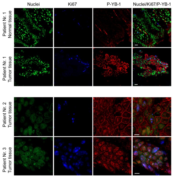
Figure 8.
YB-1 is highly phosphorylated in CRC patient tumor tissues. Paraffin-embedded sections were used for immunofluorescence staining of phospho-YB-1 in tumor tissues and the normal tissue obtained from patient no. 1. Nuclei were stained with Yopro. Ki67 was used as an indicator of cell proliferation. The sections were analyzed with a confocal laser scanning microscope. Scale bars represent 20 µm for images from patient no. 1 and 0.5 µm for patients no. 2 and no. 3.

