Figure 5.
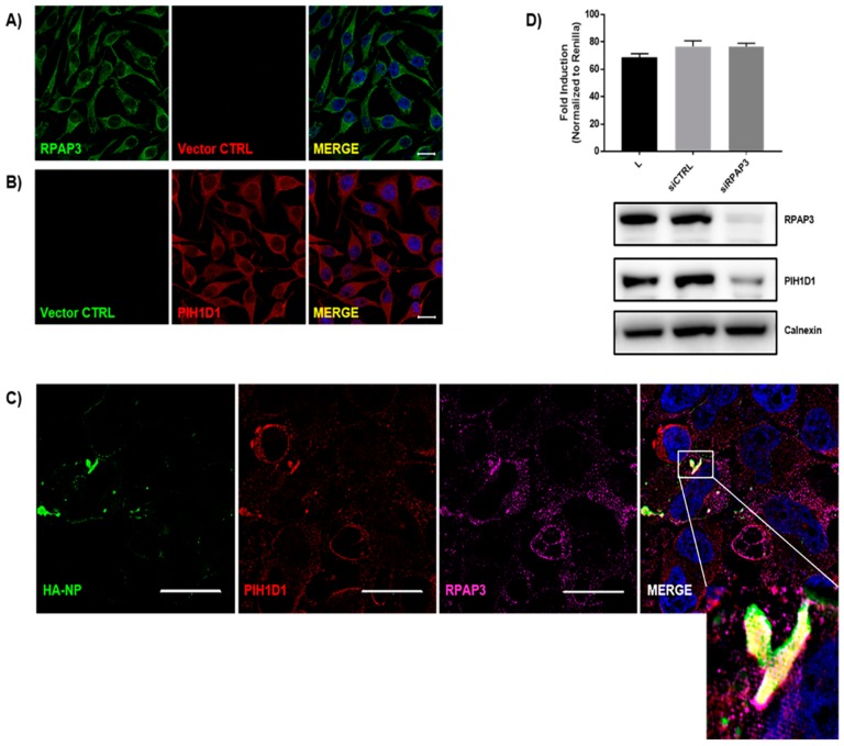
The R2TP complex components RPAP3 and PIH1D1 colocalize with HA-NP. HeLa cells were transfected with vector control or HA-NP. Twenty-four hours later, the cells were fixed and processed for the immunofluorescence detection of endogenous PIH1D1 or RPAP3 in the presence of vector control or HA-NP. Representative images of endogenous localization pattern of (A) RPAP3 with vector control and (B) PIH1D1 with vector control. RPAP3 or vector control (green), PIH1D1 or vector control (red), and Hoechst 33342 nuclear stain (blue) were visualized by confocal microscopy. Scale bars = 20 µM. (C) Representative images of endogenous localization pattern of PIH1D1 and RPAP3 in the presence of HA-NP. HA-NP (green), RPAP3 (red), PIH1D1 (magenta), and Hoechst 33342 nuclear stain (blue) were visualized by SIM. Scale bars = 20 µM. (D) Minigenome activity upon the knockdown of RPAP. Below are protein levels confirmed by immunoblot. HeLa cells were transfected with 30 nM scrambled siRNA or 30 nM siRNA targeting RPAP3. Twenty-four hours after siRNA addition, the minigenome components were transfected. Forty-eight hours later, minigenome reporter activity was measured. Data represent mean ± SEM from one representative experiment (n = 3) of at least three experiments.

