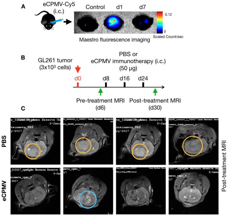
Figure 2.
Intracranial eCPMV injection and immunotherapy. (A) eCPMV retention in brain following intracranial administration was determined using eCPMV-Cy5 and Maestro fluorescence imaging system. (B) For in situ immunotherapy, C57BL6 mice (n = 4) were inoculated with 3 × 10³ GL261 cells intracranially and administered PBS or eCPMV via intracranial injections on days 8, 16 and 24. (C) On day 30, MRI imaging (7 Tesla) was used to visualize glioma post-treatment. Yellow circles highlight solid tumors in PBS administered mice, whereas blue circle highlights the residual tumor and/or edema in one of the mice in the eCPMV treatment group.

