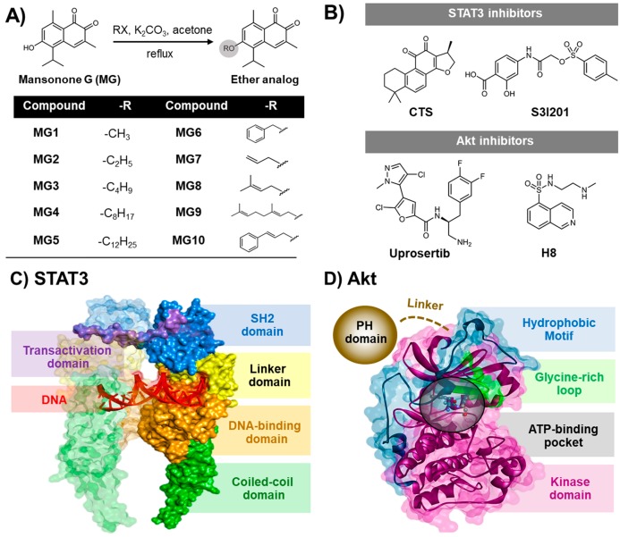
Figure 1.
Two-dimensional (2D) chemical structures of (A) MG and its semi-synthetic ether derivatives MG1-MG10 [30] and (B) the known STAT3 (cryptotanshinone (CST) and S3I201) and Akt (uprosertib and H8) inhibitors. Three-dimensional (3D) structures of (C) STAT3 and (D) Akt1 signaling proteins. The SH2 domain of STAT3 and the ATP-binding pocket of Akt are shown by blue surface and black circle, respectively.

