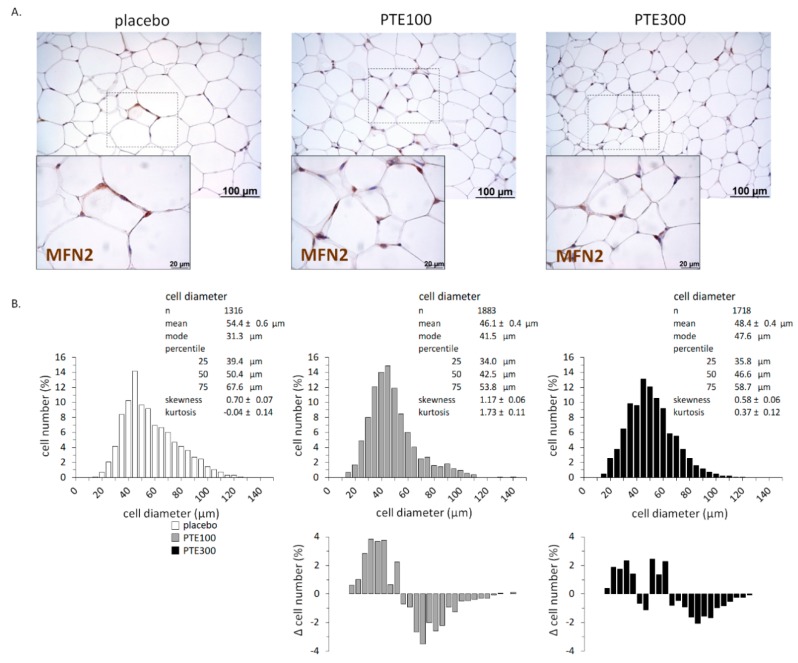
Figure 2.
Phaeodactylum tricornutum ethanolic extract (PTE) decreases adipocyte size in inguinal white adipose tissue (iWAT) of C57BL/6J mice fed with a high fat diet (HFD). Representative microphotographs illustrating adipocyte size and Mitofusin (MFN) 2 immunostaining (A), and distribution of adipocytes size (B) in iWAT at the end of the experiment. HFD-fed mice received daily an oral dose of PTE (100 mg or 300 mg/kg body weight) or placebo (olive oil:water, 2:1, v:v) for 26 days. Five to six animals per group and between 200 and 300 cells per animal were included in the analysis of distribution of adipocytes size. The area of individual adipocytes was measured using a quantitative morphometric method at 20× magnification with the assistance of Axio Vision software. Adipocyte size distribution was statistically different (p < 0.001) between the control and the PTE groups, according to the Kolmogorov–Smirnov test. The bottom panels in (B) correspond to the difference in frequency for each adipocyte size interval between the PTE-supplemented group (PTE100 or PTE300) and the control (vehicle receiving) group.

