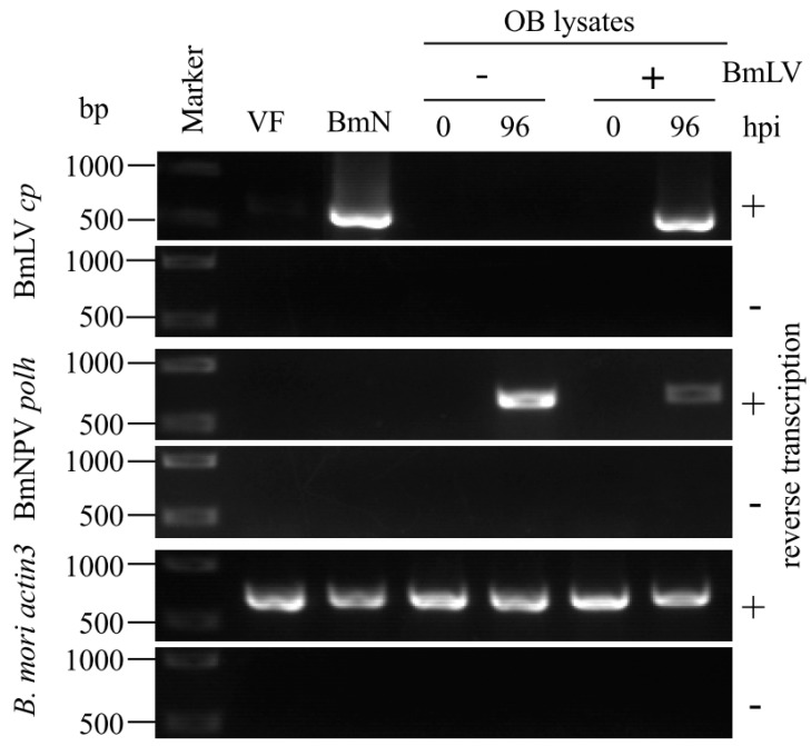
Figure 4.
Infection study of BmNPV OB lysates. BmLV-negative and -positive OBs were dissolved in an alkaline solution and inoculated onto BmVF cells. At 0 and 96 hpi, RNA was extracted and subjected to RT-PCR using primers for BmLV cp, BmNPV polh, and B. mori actin3. BmLV-negative BmVF and -positive BmN4 cells were used as controls. Size markers are indicated on the left side of the panel.

