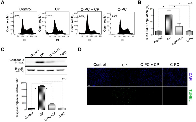
Figure 2.
Effect of C-PC on cell cycle arrest and apoptosis in cisplatin-treated HEI-OC1 cells. (A) Cell cycle analysis by flow cytometry and (B) comparison of the sub-G0/G1 ratio between the cells treated with CP alone and those pretreated with C-PC. (C) Western blot showing caspase-3 expression in cells treated with CP and C-PC. Data are shown as the mean ± standard deviation; * p < 0.05, compared with the cells treated with CP alone. C-PC, C-phycocyanin; CP, cisplatin. (D) TUNEL assay to detect apoptotic cells. Fragmented DNA (green) and nuclei (blue) were stained and observed under fluorescence microscopy. Scale bar represents 100 µm. The cells were pretreated with 1 μg/mL C-PC for 1 h, followed by treatment with 30 μM CP for 30 h.

