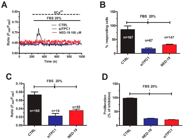
Figure 8.
TPC1 mediates FBS-induced lysosomal Ca2+ release and proliferation in mCRC cells. (A) 20% FBS induced an intracellular Ca2+ transient that was significantly reduced by NED-19 (100 μM, 30 min) and by deleting TPC1 with the specific siTPC1. (B) mean ± SE of the percentage of responding cells under the designated treatments. The asterisk indicates p < 0.05. (C) mean ± SE of the amplitude of the peak Ca2+ response to NAADP under the designated treatments. The asterisk indicates p < 0.05. (D) mean ± SE of the percentage of 20% FBS-induced cell proliferation under control conditions and upon pharmacological (NED-19) and genetic (siTPC1) blockade of NAADP-induced Ca2+ release. The asterisk indicates p < 0.05.

