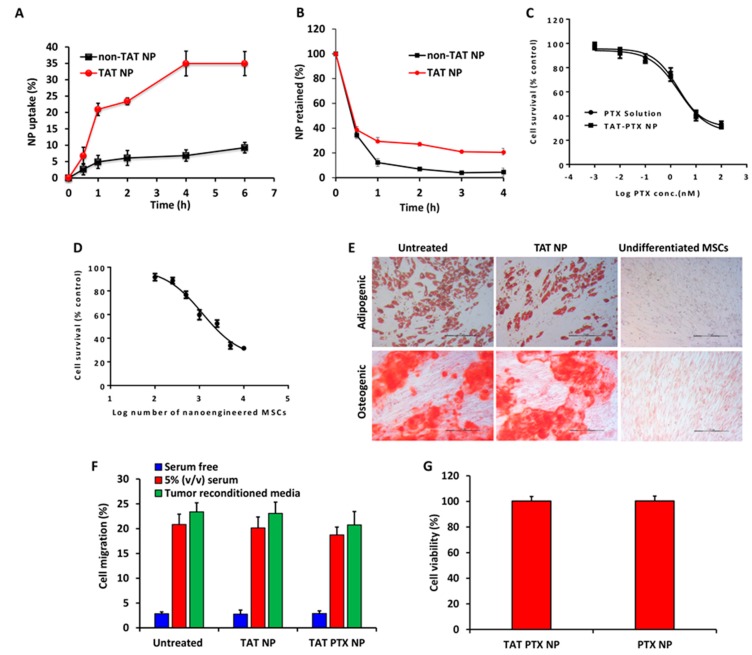
Figure 2.
In vitro endocytosis (A) and exocytosis (B) of TAT-modified or non-TAT nanoparticles in MSCs. Cells were incubated with 100 µg/mL rhodamine-labeled PLGA nanoparticles. Data shown is mean ± SD, n = 4. (C,D) Cytotoxicity profiles of PTX solution, TAT PTX NP, and nano-engineered MSCs in A549 cells. Cells were treated with PTX solution or TAT PTX NP (C) or nano-engineered MSCs (D). MTS assay was performed after 72 h of treatment. Data shown is mean ± SD, n = 6. (E) Differentiation (adipogenic and osteogenic) potential of nano-engineered MSCs (MSCs engineered with TAT NP; TAT NP). Untreated MSCs and nano-engineered MSCs were grown in adipogenic and osteogenic differentiation media for 3 weeks. The cells were fixed and stained with oil red O or alizarin red to detect lipid vacuoles or calcium deposits, respectively. MSCs cultured in regular growth media were used as negative control. Scale bar: 200 µm. (F) The migratory potential of MSCs from a serum free media towards serum free, tumor reconditioned or 5% (v/v) serum containing media in a Transwell® plate. Data shown is mean ± SD, n = 6. (G) Cell viability of nano-engineered MSCs. Data shown is mean ± SD, n = 5.

