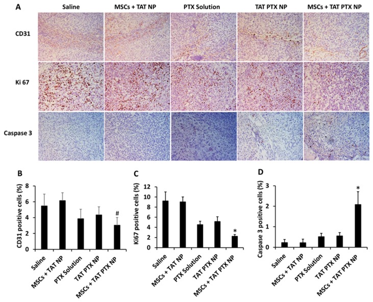
Figure 4.
Immunohistological analysis of lung tumors collected from therapeutic efficacy study. (A) Lung tumors were stained for CD31 (angiogenesis marker), Ki-67 (proliferation marker), and caspase-3 (apoptosis marker). Images were taken at 20× magnification. Quantification of (B) CD31, (C) Ki67, and (D) cleaved caspase-3 staining. Data represented as mean ± SD, n = 9 images; * p < 0.05 compared with other treatment groups and # p < 0.05 compared with saline and MSCs + TAT NP.

