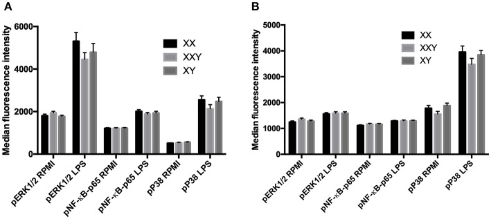
Figure 5.
Mean ± SEM of the median fluorescence intensity of phospho-ERK1/2, phospho-NF-κB-p65 and phospho-P38-MAPK in monocytes (A) and neutrophils (B), before and after stimulation of whole blood with LPS 10 μg/mL in men, women, and subjects with Klinefelter syndrome. Whole blood was stimulated with LPS at 10 μg/mL for 15 min. Reactions were stopped, and red blood cells removed. After washing, cells were permeabilized and mixed with fluorochrome-conjugated monoclonal antibodies against the phosphorylated kinases. The median fluorescence intensity of each fluorochrome was measured in each leukocyte population by flow cytometry. BD FACSDiva™ software version 6.1.2 for Windows was used to acquire the data and BD FCAP Array™ software version 1.0.1 for Windows was used to analyze the data.

