Figure 3.
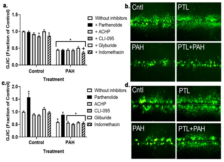
Gap junction intracellular communication (GJIC) dysregulation in response to LMW PAHs is not changed in response to anti-inflammatory inhibitors at an early time point (4 h). (a) C10 cells were treated with the binary LMW PAH mixture (40 μM; 1:1 ratio of 1-methylanthracene and fluoranthene) for 4 h following 1 h pre-incubation with these inhibitors (parthenolide, 10 μM); ACHP, 1 μM; CLI-095, 3 μM; glyburide, 50 μM; and indomethacin, 1 μM). (b) Representative images of C10 cells following the SL/DT assay used to quantify the gap junction activity in these cells in response to the PAHs and parthenolide (all other inhibitor combinations shown in Figure S1). (c) E10 cells were treated the same as the C10 cells with these inhibitors (parthenolide, ACHP, CLI-095, glyburide, and indomethacin). * p < 0.05 inhibitors or inhibitors + PAH treatment compared to control (DMSO); +, p < 0.05 indomethacin + PAH treatment compared to PAH without inhibitors (white bar on right). (d) Representative images of E10 cells following the SL/DT assay in response to the PAHs and parthenolide (all other inhibitor combinations shown in Figure S1). Experiments were repeated 3 times; n = 3 per treatment per experiment. PTL, parthenolide. Magnification 100×.

