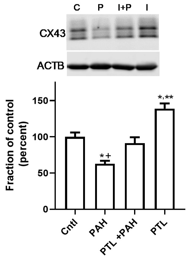
Figure 5.
CX43 protein is not suppressed by LMW PAH exposure in the C10 cells in the presence of PTL. C10 cells were pre-treated for 1 h with 10 μM parthenolide (PTL) prior to treatment with the LMW PAH mixture (15 μM; 1:1 ratio of 1-methylanthracene and fluoranthene) for 24 h. Experiment repeated 4 times; n = 3 per treatment per experiment. A representative CX43 immunoblot is depicted above the graph (* p < 0.05 for treatment compared to control), PAH treatment and PAH treatment in the presence PTL (+ p < 0.05 for PAH compared to PAH plus PTL), and PTL alone (** p < 0.05 PTL compared to control). CX43 protein expression (upper blot) is normalized to β actin (ACTB, lower blot). C, cntl, control (DMSO); P, PAH, LMW PAH mixture; I, PTL, parthenolide (inhibitor); I + P, parthenolide plus PAH treatment.

