Figure 7.

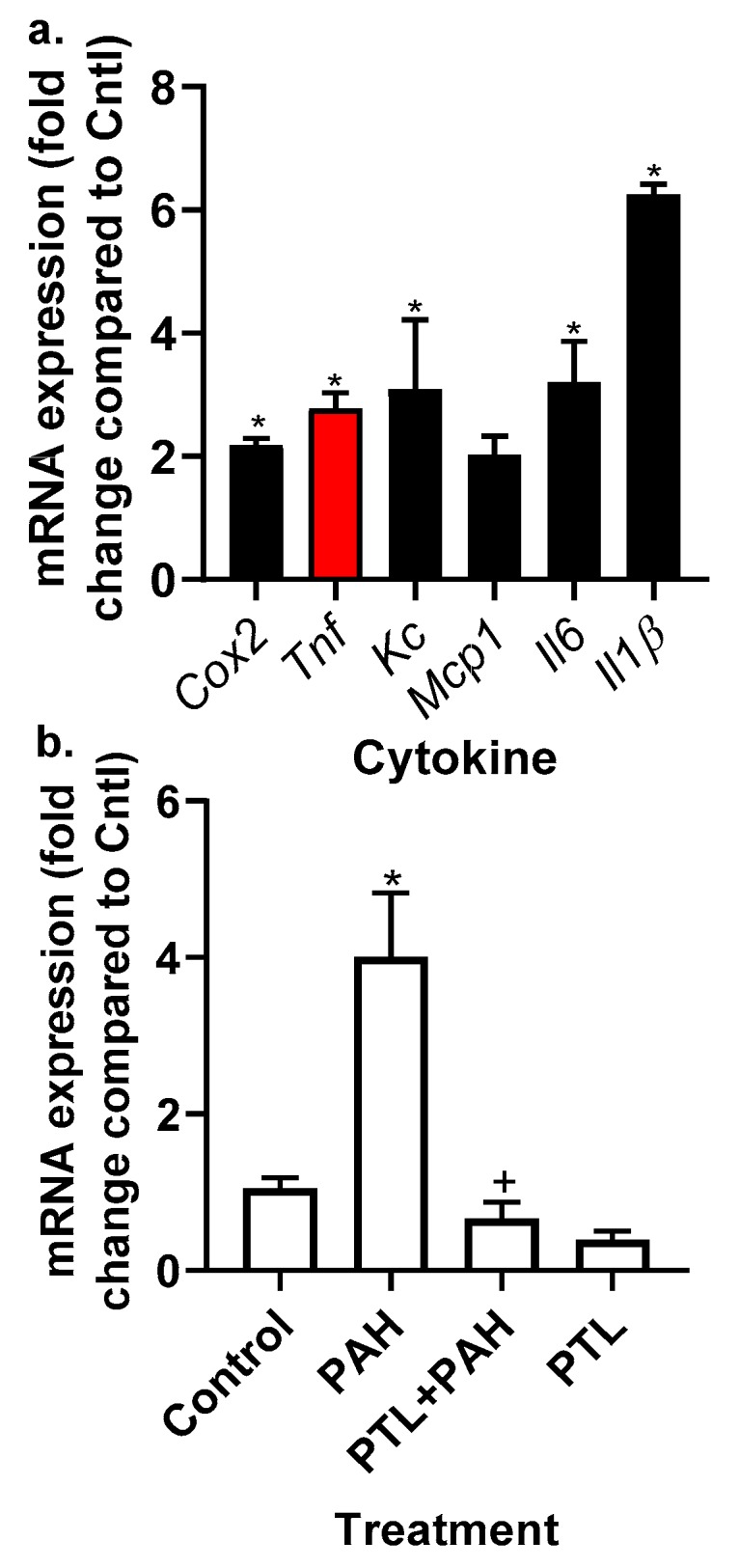
Inflammatory mediator gene expression was elevated in the MHS cells in response to the LMW PAH mixture. (a) In response to the 20 μM LMW PAH mixture (1:1 ratio of 1-methylanthracene and fluoranthene), Cox2, Tnf, Kc, Mcp1, Il6, and Il1β were all significantly elevated (* p < 0.05) compared to controls (DMSO) cells following 4 h of treatment in the MHS cells. mRNA for each cytokine gene was normalized to 18S and then fold change determined compared to control. Cox2, cyclooxygenase 2; Mcp1, monocyte chemo-attractant protein 1; IL6, interleukin 6. Tnf (red bar) indicates the cytokine we used for the recombinant cytokine studies that follow. (b) PTL inhibits Tnf expression in response to PAH exposure. * p < 0.05 PAH compared to control; + p < 0.05 PTL + PAH compared to PAH treatment. Experiments repeated 2–3 times with an n = 3 per experiment.
