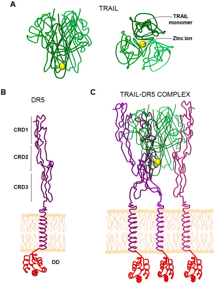
Figure 1.
Structure of (TNF)-related apoptosis-inducing ligand (TRAIL) and DR5. (A) Structure of the TRAIL trimer. Left panel corresponds to the side view and right panel to the down view. Yellow sphere corresponds to a Zinc ion. (B) Structure of the DR5 (TRAIL-R2) monomer anchored to cell surface. CRD (cystein rich domain). DD (death domain). (C) TRAIL-DR5 complex. The TRAIL trimer is drawn as tubes rendering in gradations of green, and the three receptor molecules are rendered as tubes in gradations of purple.

