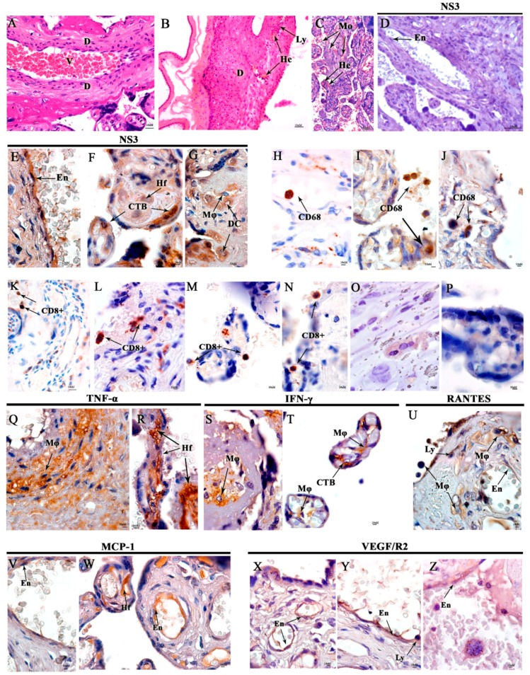
Figure 2.
Histopathological and immunohistochemistry analysis of the placenta. (A) Non-DENV patient stained with HE and presenting normal maternal decidua (D) and blood vessels (V). (B) Maternal decidua (D) from the DENV case presenting focal hemorrhage and lymphocytic infiltrates. (C) Chorionic villus with mononuclear infiltrate (Mo) and haemorrhage (He) and also intervillus space in the DENV case. (D) Placenta control with no DENV NS3 protein detection. (E) DENV NS3 protein detection in endothelial cells (En) from the maternal vessel, (F) in the cytotrophoblasts (CTB) and Hofbauer cells (Hf), (G) in decidual cells (DC), and macrophages (Mø) in the infected placenta. (H) CD68 detection in negative control and in the DENV case (I,J). (K) CD8+ T cells detection in negative control and in the DENV case (L–N). (O,P) Representative negative control of cytokine and inflammatory mediators from a non-DENV case (Q,R) Expressing TNF-α in macrophages (Mø) and Hofbauer cells (Hf) in the DENV-case. (S) Expressing IFN-y in macrophages (Mø) in maternal region, (T) in Macrophages (Mø) and cytotrophoblasts (CTB) in chorionic villi. (U) Expressing RANTES in Macrophages (Mø), endothelial cells (En), and lymphocytes (Ly) in the maternal region. (V) Endothelial cells (En) expressing MCP-1 in the maternal region and, (W) endothelial cells (En), and Hofbauer cells (Hf), both in chorionic villi. (X–Z) Macrophages (Mø), endothelial cells (En) and lymphocytes (Ly) expressing VEGF/R2.

