Figure 1.
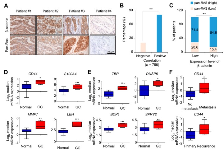
β-catenin and pan-RAS levels, and the expression of Wnt/β-catenin and RAS-ERK pathway target genes, including the CSC marker genes, were increased in tissues of GC patients. (A) Tumor tissues of 756 advanced gastric cancer (AGC) patients fixed on TMA slides were analyzed by 3,3′-diaminobenzidine staining using β-catenin or pan-RAS antibody. Representative images are presented. Scale bar represents 200 μM. (B) Correlations of the expression levels of pan-RAS and β-catenin in 756 AGC tissues are presented as percentages (P < 0.01). (C) Correlations of the expression levels of pan-RAS and β-catenin are presented on the basis of high and low patterns of these markers (P < 0.01, chi-squared test; comparing β-catenin low versus high). (D–E) Oncomine analysis of the DErrico database for the mRNA expression of Wnt/β-catenin (CD44, S100A4, MMP7, and LBH) and RAS-ERK (TBP, DUSP6, BDP1, and SPRY2) pathway target genes in GC tissues compared with normal gastric mucosa tissues (n = 69, DErrico Gastric statistics; P < 0.05 with Student’s t-test). (F) Oncomine analysis for the mRNA expression of CD44 in metastatic and recurring GC patient tissues compared with non-metastatic and primary tumor tissues, respectively (n = 43, Forster Gastric statistics; P < 0.05 with Student’s t-test).

