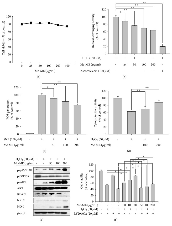
Figure 1.
Antioxidative and cytoprotective effects of Mc-ME in HaCaT cells. (a) HaCaT cells were treated with increasing doses of Mc-ME (0–400 μg/ml) for 24 h, and cell viability was determined by a conventional MTT assay. (b) DPPH (150 μM) was mixed with either increasing doses of Mc-ME (0–200) or ascorbic acid (100 μM) and incubated for 20 min. The radical scavenging activity was determined by measuring the absorbance at 517 nm. (c) HaCaT cells were pretreated with increasing doses of Mc-ME (0–200 μg/ml) for 30 min and then treated with SNP (200 μM) for 24 h. The cells were incubated with H2DCFDA (10 μM) at 37°C for 20 min, and ROS levels were determined by measuring fluorescence using a flow cytometer. (d) HaCaT cells were pretreated with increasing doses of Mc-ME (0–200 μg/ml) and then treated with H2O2 (50 mM) for 24 h. Cell viability was determined by a conventional MTT assay. (e) HaCaT cells were pretreated with increasing doses of Mc-ME (0–200 μg/ml) and then treated with H2O2 (50 μM) for 24 h. Levels of total and phosphorylated KEAP1, HO-1, p85/PI3K, and AKT in the total cell lysates were determined by western blot analysis. (f) HaCaT cells were treated with H2O2 (50 mM) and/or LY294002 (20 μM) in the absence or presence of increasing doses of Mc-ME (0–200 μg/ml) for 24 h and cell viability was determined by a conventional MTT assay. ∗P < 0.05, ∗∗P < 0.01 compared to control.

