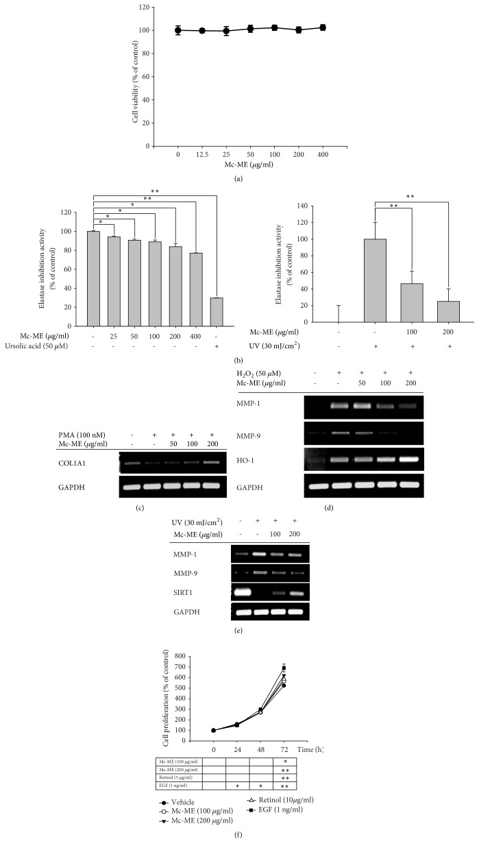
Figure 2.
Regulatory effect of Mc-ME on skin tissue remodeling factors in NIH3T3 and HaCaT cells. (a) NIH3T3 cells were treated with increasing doses of Mc-ME (0–400 μg/ml) for 24 h, and the cell viability was determined by a conventional MTT assay. ((b); left panel) Elastase (0.3 units/ml) and STANA (5 mM) were mixed with either increasing doses of Mc-ME (0–400 μg/ml) or ursolic acid (50 μM), followed by incubation for 15 min. ((b); right panel) NIH3T3 cells were irradiated at 30 mJ/cm2 for 10 sec in the absence or presence of increasing doses of Mc-ME (0–200 μg/ml) followed by incubation for 24 h. Cell lysates were incubated with elastase (0.3 units/ml) and STANA (5 mM) for 15 min. Elastase activity was determined by measuring the absorbance at 410 nm. (c) HaCaT cells were pretreated with increasing doses of Mc-ME (0–200 μg/ml) for 30 min and then treated with PMA (100 nM) for 24 h. mRNA expression level of COL1A1 was determined by semiquantitative RT-PCR. (d) HaCaT cells were pretreated with increasing doses of Mc-ME (0–200 μg/ml) for 30 min and then treated with H2O2 (50 μM) for 24 h. mRNA expression levels of MMP-1, -9, and HO-1 were determined by semiquantitative RT-PCR. (e) HaCaT cells were irradiated at 30 mJ/cm2 for 10 sec in the absence or presence of increasing doses of Mc-ME (0–200 μg/ml), and mRNA expression levels of MMP-1, -9, and SIRT1 were determined by semiquantitative RT-PCR. (f) HaCaT cells were treated with increasing doses of Mc-ME (0–200 μg/ml), RE (10 μg/ml), or EGF (1ng/ml) for 72 h, and the cell proliferation level was determined by measuring cell viability with a conventional MTT assay every 24 h. ∗P < 0.05, ∗∗P < 0.01 compared to control.

