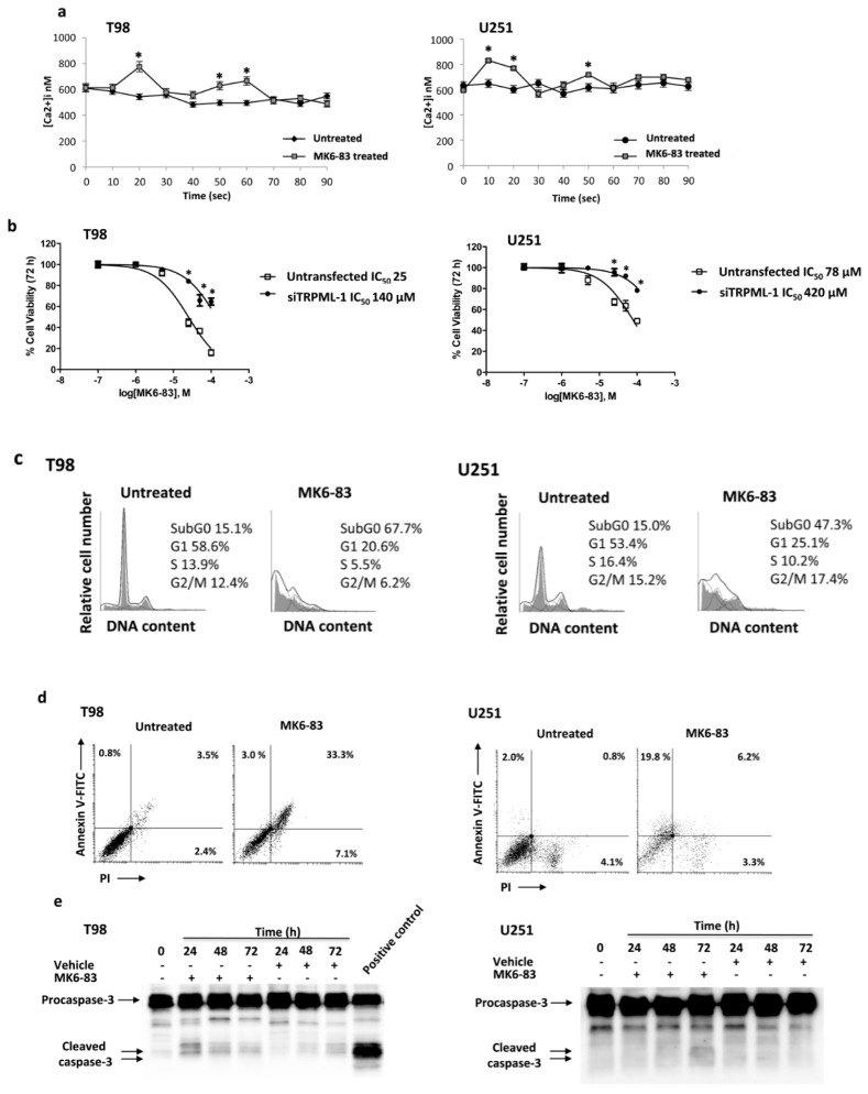
Figure 4.
MK6-83 induces TRPML-1 activation and triggers T98 and U251 apoptotic cell death. (a) Time course of the [Ca2+]i rise was evaluated by FACS analysis in T98 and U251 GBM cells untreated or treated with 10 μM and 25 μM of MK6-83, respectively. Data shown are the mean ± SD of three independent experiments. Statistical analysis was determined by comparing MK6-83-treated with untreated cells, * p < 0.05. (b) Cell viability was evaluated by 3-(4,5-dimethylthiazol-2-yl)-2,5-diphenyltetrazolium bromide (MTT) assay in untransfected or TRPML-1-silenced (siTRPML-1) T98 and U251 GBM cells treated with different doses of MK6-83 for 72 h. Data shown are expressed as mean ± SE of three separate experiments. (c) Representative cell cycle distribution in GBM cells treated for 72 h with MK6-83 10 μM in T98 and 25 μM in U251 cells. Data are one out of three separate experiments. (d) Biparametric flow cytometric analysis was performed in T98 and U251 cells, untreated or treated with MK6-83 for 48 h, by Annexin V- Fluorescein isothiocyanate (FITC) and Propidium iodide (PI) staining. Cells in the upper left quadrant indicate Annexin V-positive, early apoptotic cells. The cells in the upper right quadrant indicate Annexin V-positive/PI-positive, late apoptotic cells. (e) Lysates from T98 and U251 cells, untreated or treated with MK6-83 for different times, and from positive control for caspase-3 activation were separated on SDS-PAGE and probed with anti-caspase-3 Ab. Blots are representative of three separate experiments.

