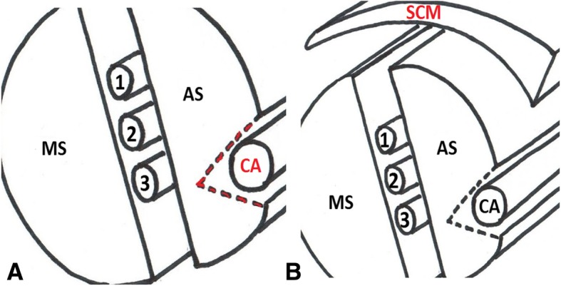
Fig. 6.

Incision in red indicates the location of the carotid artery (a). Thin layer of meat covering the model simulates the sternocleidomastoid muscle (b). SCM, sternocleidomastoid muscle; AS, anterior scalene muscle; MS, middle scalene muscle; CA, carotid artery; the interscalene brachial plexus is located between AS and MS muscles; 1, C5 nerve root; 2, C6 nerve root; 3, C7 nerve root
