Fig. 1.

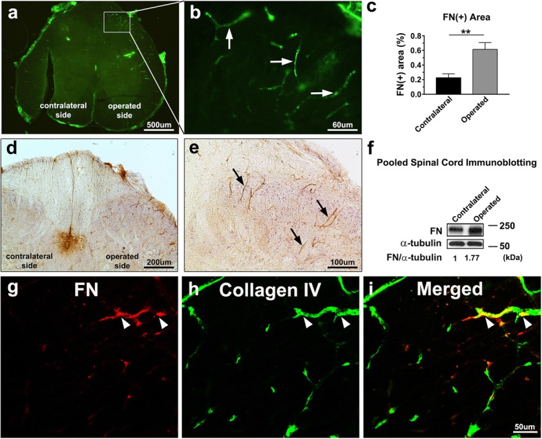
Spinal nerve ligation induces excessive FN deposition around microvessels in the spinal cord. The L5 spinal nerve distal to the dorsal root ganglion of rats was tightly ligated. a Representative photomicrograph depicting immunofluorescence analysis for FN on the operated and contralateral sides of the L5 segment of the spinal cord 7 days after surgery. b Regional magnification of the boxed area in (a); FN+ microvessel-like profiles are indicated by arrows. c Percentages of the FN+ area in the total area on the operated and contralateral sides of the L5 segment of the spinal cord were analyzed (n = 3). Data are presented as means ± standard errors of means. **P < 0.01, Student’s t test. d and e Representative images at low (d) and high (e) magnification showing immunocytochemistry of FN in the L5 dorsal part of the spinal cord. Arrows indicate FN+-microvessel-like profiles on the operated side of the spinal cord. f Immunoblotting for FN expression in the pooled L5 dorsal spinal cord on the operated and contralateral sides in five male Sprague Dawley rats. Equal protein loading was confirmed with α-tubulin. Quantification of immunoblotting of FN normalized to α-tubulin in tissues is shown. g–i Confocal microscopic images of FN+-microvessel-like profiles (red; g) and collagen IV+ capillaries (green; h) in the L5 dorsal part of the spinal cord; merged images (i) showing the colocalization of FN and collagen IV (yellow) in the capillaries are indicated with arrowheads
