Fig. 2.

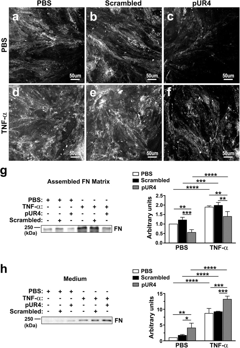
TNF-α-induced FN deposition in bEND.3 cells is inhibited by pUR4. After being incubated with 1000 nM scrambled peptide, 1000 nM pUR4, or PBS for 16 h, bEND.3 cells underwent treatment with TNF-α (20 ng/mL) or PBS for 24 h. a–f Immunofluorescence analysis for FN was performed on nonpermeabilized cells to evaluate FN fibrils accumulated outside bEND.3 cells. g–h To assess the extracellular matrix FN deposited by bEND.3 cells after the various treatments, cellular components were extracted using the lysis buffer, as described in the Methods section, and the assembled ECM FN by bEND.3 cells (g) and FN in the corresponding conditioned medium (h) were immunoblotted. Quantification of protein band intensity was determined (n = 3). Data are presented as means ± standard deviations. *P < 0.05, **P < 0.01, ***P < 0.001, and ****P < 0.0001, two-way ANOVA followed by Tukey’s multiple comparison test and Sidak’s multiple comparison test
