Fig. 6.

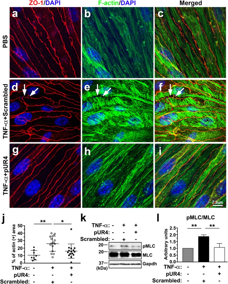
TNF-α-induced stress fiber formation and MLC phosphorylation are attenuated by pUR4. After being pretreated with 1000 nM scrambled peptide or 1000 nM pUR4 for 16 h, bEND.3 cells were stimulated with 20 ng/mL TNF-α for 24 and 6 h. a–i Cytoskeletal remodeling in response to TNF-α treatment for 24 h was analyzed through immunofluorescence analysis for ZO-1 (red) and phalloidin staining for F-actin (green) in PBS control and TNF-α-treated bEND.3 cells. Arrows indicate paracellular gaps. j Quantitative analysis of the F-actin+ area in the total area in PBS control and TNF-α-treated bEND.3 cells with scrambled peptide or pUR4 preincubation (n = 3). Data are expressed as means ± standard deviations. *P < 0.05 and **P < 0.01 by one-way ANOVA followed by Tukey’s multiple comparison test. k and l After exposure to TNF-α for 6 h, MLC phosphorylation was examined by immunoblotting with phospho-MLC (T18/S19)-specific antibody (pMLC), and total MLC (MLC) antibody on the same membrane after stripping (k). Quantification of immunoblotting of pMLC normalized to MLC in the bEND.3 cells (l; n = 3). Data are represented as means ± standard deviations. **P < 0.01, one-way ANOVA followed by Tukey’s multiple comparison test
