Fig. 7.

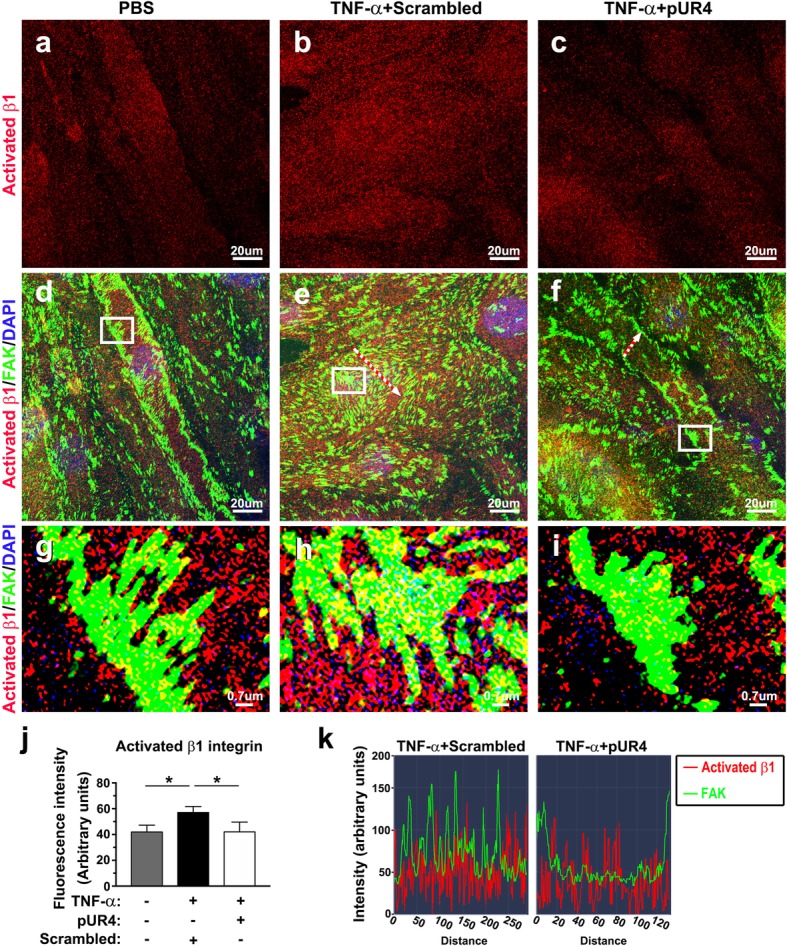
TNF-α-induced β1 integrin activation and clustering to focal adhesions (FAs) are diminished by pUR4. After being pretreated with 1000 nM scrambled peptide or 1000 nM pUR4 for 16 h, bEND.3 cells were stimulated with TNF-α (20 ng/mL) for 24 h. PBS-treated control and TNF-α-treated bEND.3 cells were costained with antibodies to the activated state of β1 integrin (red) and FAK (green). Three independent experiments were performed, and each experiment was repeated with similar results. a–c Representative immunofluorescence images showing activated β1 integrin in control and bEND.3 cells treated with the scrambled peptide and pUR4.d–f The merged images show colocalization (yellow) of activated state of β1 integrin (red) and FAK (green). g, h, and i Reginal enlargement of the FAs in the boxed area in (d), (e), and (f), respectively. (j) Quantification of FI of activated β1 integrin in PBS control and bEND.3 cells treated with the scrambled peptide and pUR4 was derived from a representative experiment (n = 3). Data are represented as means ± standard deviations. *P < 0.05, one-way ANOVA followed by Tukey’s multiple comparison test. (k) Line scan graphs showing the immunofluorescence intensity along the freely positioned arrows in bEND.3 cells treated with scrambled peptide (e) and pUR4 (f)
