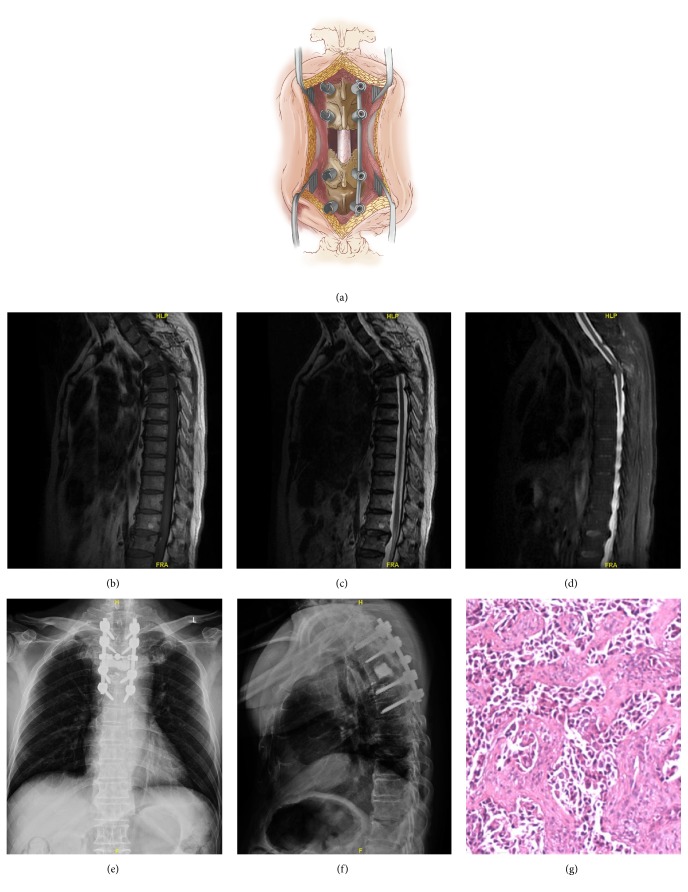
Figure 2.
Case example of a 62-year-old male with metastatic lung cancer and walking difficulty. Illustrations showing the open approach, demonstrating the fascia and muscle opened at every level of the exposed region (a). Metastatic epidural spinal cord compression of T4 is seen on thoracic magnetic resonance imaging (b-d). The patient underwent laminectomy, corpectomy, tumor resection, and polymethylmethacrylate reconstruction of T4, plus pedicle screw instrumentation of T2-T6. Postoperative imaging shows stable reconstruction (e-f). Pathological examination shows that the lesion was consistent with adenocarcinoma of the lung (g).

