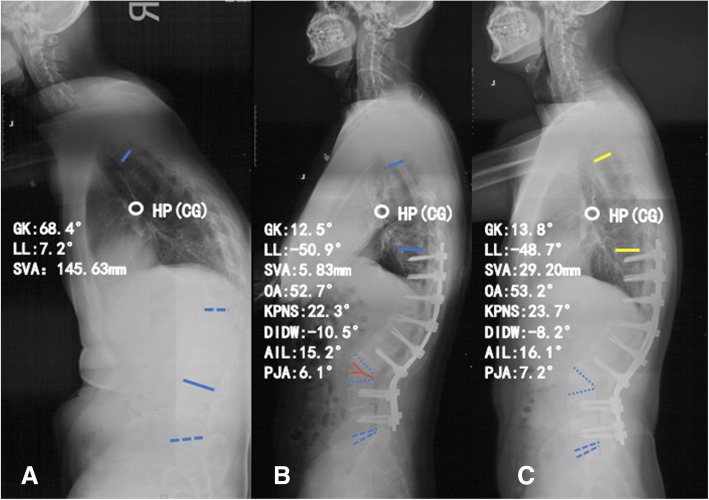
Fig. 3.
A 38-year-old male patient with thoracolumbar kyphosis secondary to ankylosing spondylitis. a Preoperative standing lateral radiograph. b Immediately after surgery with vertebral column decancellation (VCD) technique. c Lateral radiograph taken 32 months after surgery. AIL angle of instrumented levels, DIDW the sum of distal non-fused intervertebral disc wedging, GK global kyphosis, KPNS kyphotic angle of proximal non-fused segment involved in the global kyphosis, LL lumbar lordosis, OA osteotomized vertebra angle, PJA proximal junctional angle, SVA sagittal vertical axis, HP hilus pulmonis, CG the center of gravity

