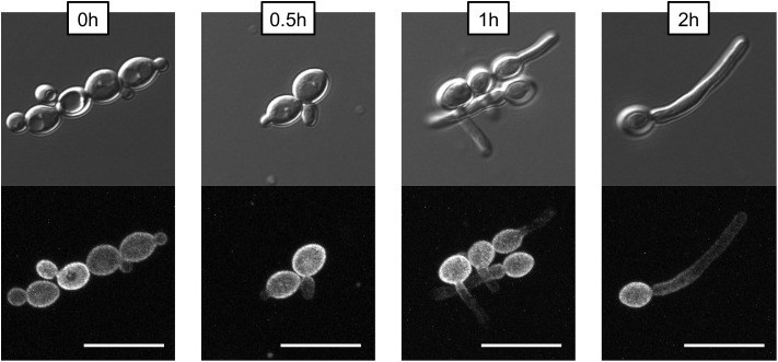
FIGURE 9.
Non-homogenous distribution of Pma1p in hyphae. A wild-type strain in which PMA1 was C-terminally tagged with GFP was diluted to a concentration of 5 × 10⁶ cells per mL and grown in YNB+20% FCS for 4 h. Cells were imaged using a confocal microscope to demonstrate that hyphal tips are not simply out of the field of view. For the GFP fluorescent image, a Z-stack comprised of 0.2-micron thick slices is shown. Pma1-GFP is distributed evenly across the plasma membrane in yeast cells, but is less prominent along the hyphae of filamenting cells. Scale bars indicate 10 μm.

