FIGURE 5.
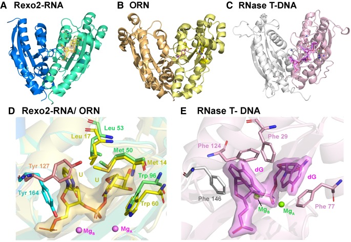
The crystal structure of Rexo2 strongly resembles that of ORN. (A) Crystal structure of Rexo2–RNA with the active-site residues and RNA shown in stick model. (B) Crystal structure of the apo form of ORN (PDBID :2IGI) from E. coli. (C) Crystal structure of RNase T bound with DNA (PDBID: 3NH1). (D) Superimposition of RNA-binding aromatic residues in the active site of Rexo2–RNA and ORN revealing that these residues are located at similar positions. (E) Stick representation of the two 3′-end DNA nucleobases (dGdG) that make π–π stacking interactions with the four aromatic Phe residues in the active site of RNase T.

