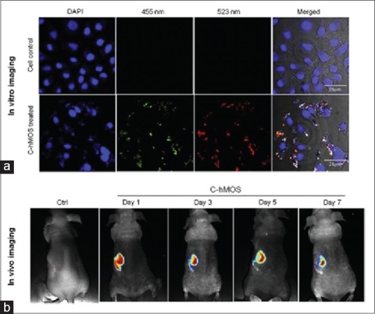
Figure 3.

(a) Images of treated cells by 40 μg/ml dose of created mesoporous hollow organosilica (C-hMOS) nanoparticles, after a period of 4 h. To visualize nanoparticles inside cells, different wavelength used.(b) in vivo images of nude mouse which treated by C-hMOS and monitored for 7 days[63]
