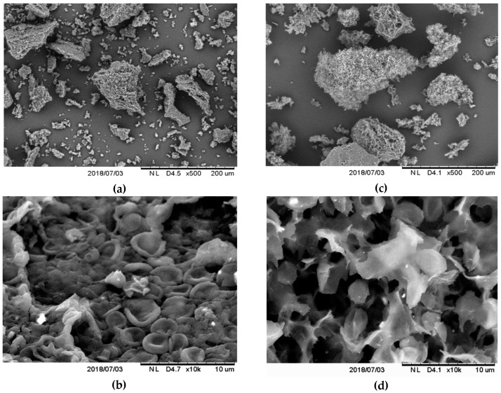
Figure 1.
Examples of microscopic structure of studied cell walls (CW) and purified β-glucan (β-G) particles of C. utilis ATCC 9950 obtained by scanning electron microscopy at different magnifications (D—working distance from electron source); (a,b) morphology of CW particles; (c,d) morphology of purified β-G particles.

