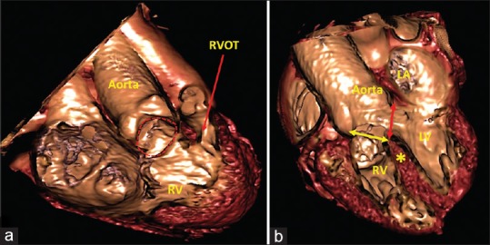
Figure 4.

Virtual cardiac dissection from an adolescent boy with Tetralogy of Fallot. (a) An image in right anterior oblique projection with cranial angulation shows the ventricular septal defect (red dotted line) and narrowing of the right ventricular outflow tract. The relationship of the ventricular septal defect with the tricuspid valve apparatus is also well seen. (b) Equivalent to fluoroscopic left anterior oblique projection with cranial angulation shows a large subaortic ventricular septal defect with overriding of the aortic root over the crest of the muscular septum (yellow asterisk). Surgeons will have to close the defect at its right ventricular margin (yellow double-headed arrow) to connect aorta to the left ventricle. The left ventricular margin (red double-headed arrow) is the outflow tract for the left ventricle, and can never be closed. LV: Left ventricle, RV: Right ventricle
